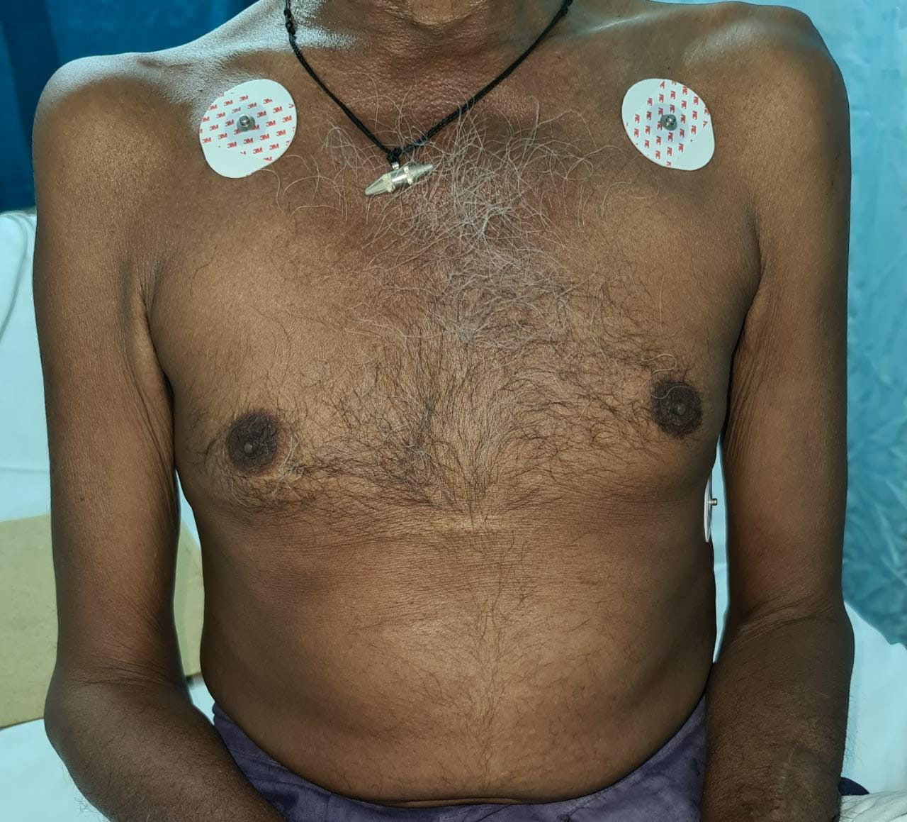
BIMONTHLY ASSESSMENT FOR THE MONTH -FEBRUARY
BIMONTHLY ASSESSMENT FOR THE MONTH OF FEBRUARY :
This is my submission for the Bimonthly internal assessment for the month of February."
The questions to the cases being discussed can be viewed from the link below
https://medicinedepartment.blogspot.com/2021/02/medicine-paper-for-february-2021.html?m=1
1Q. 50 year man, he presented with the complaints of
Frequently walking into objects along with frequent falls since 1.5 years
Drooping of eyelids since 1.5 years
Involuntary movements of hands since 1.5 years
Talking to self since 1.5 years
a. What is the problem representation of this patient and what is the anatomical localization for his current problem based on the clinical findings?
This was our unit case came to OPD on 10/2/21.
I was read about this case but it's first time for me to see such rare case.
Coming to the case,
He was 50yr old who was diagnosed as diabetic 9 months back came with complaints of
Frequently walking into objects along with frequent falls since 1.5 years
Drooping of eyelids since 1.5 years
Involuntary movements of hands since 1.5 years
Talking to self since 1.5 years
Bed wetting since 1 year
He had reduced arm swing
H/O suicidal attempt present.
ANATOMICAL LOCALIZATION TO HIS PROBLEMS:
* Drooping eyelids ...oculomotor nerve is the nerve supply . It's arises from midbrain
*Involuntary movements and frequent falls
Suggests of Basal Ganglia
*Self talking and bed wetting- frontal lobe (Pre frontal area Broadmann 8,9)
*Suicidal attempt - ?involvement of limbic system (temporal lobe area)
AS MULTIPLE AREAS OF BRAIN WERE INVOLVED I WOULD PROBABLY GO IN FAVOUR OF NEURODEGENERATIVE DISORDER
b) What is the etiology of the current problem and how would you as a member of the treating team arrive at a diagnosis? Please chart out the sequence of events timeline between the manifestations of each of his problems and current outcomes.
I came over two differential diagnosis for the current case :
1.PROGRESSIVE SUPRANUCLEAR PALSY (PARKINSON PLUS SYNDROME)
Superior gaze palsy,frequent walking into objects and frequent falls,drooping of eyelids goes in favour
The important Differential is
2.MYASTHENIA GRAVIS .
It is ruled out after performing an ICE PACK Test.
There is no significant improvement of ptosis
And also there is no progressive weakness as the day progresses which is a characteristic of MG.
--)Seizures 10 years back
--)Type 2DM 2 years back
--)Sudden blurring of vision while riding bike met with RTA -- fracture in left leg ,operated 2 years back
--)Frequently walking into objects along with frequent falls,drooping of eyelids,Involuntary movements of hands,Talking to self 1.5 years backS
--)Stopped alcohol & tobacco consumption 1 year back
--)Non productive cough 8 months back
--)Non healing ulcer at surgical site 7 months back
--)for 1 week - diagnosed as PSP & discharged with SYNDOPA 110 MG & QUETIAPINE
--)5 days later patient presented to casualty in a state of unresponsiveness with GCS: 3/15 with H/o 2-3 episodes vomiting.
--)Another 2 episodes of generalized tonic seizures in casualty - treated with levipil
--)Suddenly his saturations & heart rate dropped with no peripheral pulsations and patient was intubated - CPR done and was resuscitated.
--)Currently on mechanical ventilator on cpap
a) What is the problem representation of this patient and wht is the anatomical localization for his current problem based on the clinical findings?
Problem representation:
A 60 year old man with a history of CVA 6 months back presented with
Dyspnea since 2 months
Bilateral pedal edema since 2 months
Reduced urine output since 2 months
Generalised weakness since 2 months
His examination findings were Visible apical impulse, Pericardial bulge, visible pulsations, dilated veinsshift of apex beat to 6th ICS, Thrill at the apex, Loud S1 present, loud P2 present, S3 Accentuating on inspiration- RVS3, Expiration - LVS3
His Ecg shows poor R wave progression
Chest Xray PA shows Cardiomegaly
His 2Echo is suggestive of Heart failure DCMP with Hypokinesia at RCA, LCX
Anatomical diagnosis:
The location to his problems is at the Heart, secondary to atherosclerosis of the vessels
a) What is the problem representation of this patient and what is the anatomical localization for his current problem based on the clinical findings?
Problem representation:
A 60 year old man with a history of CVA 6 months back presented with
Dyspnea since 2 months
Bilateral pedal edema since 2 months
Reduced urine output since 2 months
Generalised weakness since 2 months
His examination findings were Visible apical impulse, Pericardial bulge, visible pulsations, dilated veinsshift of apex beat to 6th ICS, Thrill at the apex, Loud S1 present, loud P2 present, S3 Accentuating on inspiration- RVS3, Expiration - LVS3
His Ecg shows poor R wave progression
Chest Xray PA shows Cardiomegaly
His 2Echo is suggestive of Heart failure DCMP with Hypokinesia at RCA, LCX
Anatomical diagnosis:
The location to his problems is at the Heart, secondary to atherosclerosis of the vessels
Risk factors:
Alcohol
Age of 60 years
Male gender
Etiology to his current problems :
CAD leading to DCMP
Diagnosis:
DCMP with an EF of 34% secondary to CAD
CVA 6 months back (? Left ischaemic stroke)
Benfomet as thiamine replacement in alcoholic patients
a) What is the problem representation of this patient and what is the anatomical localization for his current problem based on the clinical findings?
Problem representation:
A 52 year old man, who is a known to be a Diabetic and hypertensive presented with:
Dyspnea since 10 days
Productive cough since 2 days
Disturbed sleep since 10 days
Anatomical localization:
The anatomical location of the problem is in the lungs
Lower respiratory tract infection
b) What is the etiology of the current problem and how would you as a member of the treating team arrive at a diagnosis? Please chart out the sequence of events timeline between the manifestations of each of his problems and current outcomes.
No respiratory examination has been mentioned in the elog.
However his problems list are:
- Hyponatremia
-Lower respiratory tract infection
-Uncontrolled blood sugars
-Dimorphic Anemia
2. A 41 year man with Pancreatic pseudocyst
3. 73 year man with CAD showing a bifascicular block on the ECG posted for left inguinal hernia surgery
4. 50 year old man with HFrEF
5. 45 year man with Fever and Rash


Comments
Post a Comment